Participating in experiments at the CSLB
What is MRI scanning?
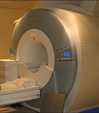 |
MRI stands for 'Magnetic Resonance Imaging'. MRI is a technique that allows us to examine in detail the structures of the brain. The basis for MRI is the use of magnetic fields and radio frequencies to produce a map of the water concentrations in the body. Because different types of tissue have different water concentrations MRI enables us to visualise where different types of tissue are located. The MRI scan involves no needles or radiation, but is noisy and may be inappropriate for people who suffer from claustrophobia, as it involves lying inside of a narrow tube during the scan.
What is fMRI scanning?
fMRI stands for 'functional Magnetic Resonance Imaging'. fMRI allows us to determine which parts of the brain are more active while performing certain tasks than while performing others. As suggested by its name, fMRI is a kind of MRI.
What will I have to do during MRI / fMRI?
Before you have your scan there will be a quick assessment to make sure that you are suitable to be scanned (a mini version of some of the questions we ask you in the Research Participant Form that you will already have completed). The scanning process will be explained to you in detail again on the day. The radiographer will ask you to lie down in the scanner and will make you comfortable. You will be given earplugs and ear defenders to block out some of the sound of the scanner. The radiographers will talk to you during the scan to check you are still comfortable and happy to continue and you will be given a squeeze ball which you can use to attract the attention of the radiographer at any time. During the first part of the fMRI scan we will take a short series of images to position your brain. You will then be asked to perform simple tasks such as reading words and making decisions about those words by pressing a button, or passively listening to words. During the later part of the fMRI scan you will simply be lying still while the scanner acquires a detailed map of your brain. You will be inside the scanner for approximately one hour or so, and the entire process will last around 2 hours from the time you arrive until you leave.
What is MEG scanning?
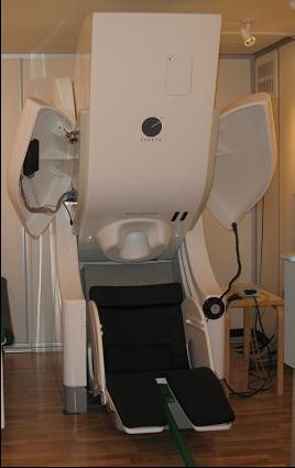 |
MEG stands for 'Magnetoencephalogram'. This records the small changes in magnetic fields at the surface of the head which are naturally generated by nerve cell activity inside the brain. You are not exposed to external electrical or magnetic fields. The top of your head will simply be positioned within a helmet-like device that can measure the very weak magnetic fields produced as a normal consequence of activity in your brain. A handful of sensors will also be attached to your forehead with tape (which should not cause discomfort nor leave any mark) to measure the precise position of your head relative to the helmet and to record movement such as blinking. Because the magnetic signals we measure are very weak, we would like to avoid any possible source of recording artefacts. In particular, it is important that muscle activity, eye movements and eye blinks are minimised (though, of course, not completely suppressed). We will therefore ask you to sit as still as possible during the experiment.
What will I have to do during MEG?
Before you have your scan, there will be a quick assessment to make sure that you are suitable to be scanned (a mini version of some of the questions we ask you in the Research Participant Form that you will already have completed). The procedure that you are about to undergo will once again be explained to you in detail. The MEG operator will ask you to sit comfortably in the scanner in a relaxed position. During a typical experiment, for example, you might be presented with visual words or pictures on a screen in front of you, or hear sounds or words through headphones, and may be asked to press buttons with your fingers according to a simple task. The precise experimental procedures will of course be explained in more detail and you will be given the opportunity to practice and ask questions. You will be inside the scanner for approximately one hour or so, and the entire process will last around 2 hours from the time you arrive until you leave.
What is EEG scanning?
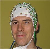 |
EEG stands for 'Electroencephalography'. This uses electrodes fitted into a plastic cap which records the electrical currents on the surface of your head which are naturally generated by the brain. You are not exposed to external electrical or magnetic fields.
The cap takes approximately 1-1¼ hours to fit. We will ask you to sit in a chair while we fit the cap, making sure each of the electrodes is in contact with your head. Once the cap is fitted, we add a conductive gel to each electrode to improve the quality of the signal we get. This gel is cool and wet and so you will probably want to wash your hair after the scan. We will also attach some sensors to your forehead with removable tape which are used to monitor your eye movements.
EEG scanning can be done on its own, or in combination with MEG scanning. If we are collecting EEG data at the same time as MEG data, you will go into the MEG scanner after the EEG cap has been fitted.
Because the electrical signals we measure are very weak, we would like to avoid any possible source of recording artefacts. In particular, it is important that muscle activity, eye movements and eye blinks are minimised (though, of course, not completely suppressed). We will therefore ask you to sit as still as possible during the experiment.
What will you have to do during EEG?
Before you have your scan, there will be a quick assessment to make sure that you are suitable to be scanned (a mini version of some of the questions we ask you in the Research Participant Form that you will already have completed). The procedure that you are about to undergo will once again be explained to you in detail. The researcher will take time to correctly fit the EEG cap and apply the conductive gel. If you are having EEG at the same time as MEG, we will ask you to sit in the MEG scanner in a comfortable position. During a typical experiment, for example, you might be presented with visual words or pictures on a screen in front of you, or hear sounds or words through headphones, and may be asked to press buttons with your fingers according to a simple task. The precise experimental procedures will of course be explained in more detail and you will be given the opportunity to practice and ask questions. You will be asked to keep as still as possible but there will be the opportunity to take breaks. The EEG scan will last approximately one hour or so, and the entire process will last around 2-3 hours from the time you arrive until you leave.
Other frequently asked questions
Who cannot take part in neuroimaging studies?
Because of the strong magnetic fields used in the MRI scanner and the magnetic fields we measure in MEG, people with implants such as cardiac pacemakers, aneurysm clips in their brain, cochlear implants and permanent eyelining cannot take part. Most types of metallic implant are not suitable but some, such as artificial joints, small pins/screws in bones, and some dental work may be suitable. Anyone who has spent significant amounts of time doing metal work involving a lathe, grinder, or similar tool should not take part. For MEG and EEG, you must not have dyed your hair in the two weeks before the experiment. People who are currently taking drugs that affect mental activity, such as tranquillisers, sleeping pills, or anti-depressants cannot take part in these studies. People with a history of brain damage or psychiatric intervention may not be able to participate.
Both men and women over 18 years of age are encouraged to participate. The narrow space inside the fMRI scanner may be a problem for some people, even for those with no history of claustrophobia. If you experience any discomfort during the scan or would like the scan to stop you can alert the radiographers at any time using the alarm you are given.
The MEG scanner is more spacious as only the top and sides of the head are covered but those with claustrophobia may still find it difficult and therefore may not wish to participate. Again, if you are in any discomfort you can make the MEG operator aware of this and we will stop the scan.
What are the positive aspects for volunteers?
When you volunteer to undergo scanning, you can go away with the knowledge that the experience has contributed to furthering the progress of medical and psychological research. You will receive £10/hour and we will also give you a copy of your MRI scan when you leave so that you can have a picture of your brain to keep!
Will a doctor look at my MRI scan?
Like faces, brains come in all shapes and sizes, so that there are many normal variations of what the scan shows. There is a very small chance that your scan may show a significant abnormality of which you are unaware. In such circumstances, you will be appropriately counselled. You will be referred to the appropriate specialist in consultation with your General Practitioner if that is what you would like. Such early detection has the benefit of starting treatment early but, in a small number of cases, may have implications for future employment and insurance.
Who can I contact if I have further questions?
For questions regarding particular experiments conducted by the Centre for Speech, Language and the Brain please contact:
Kathy Purdy
Centre for Speech, Language and the Brain
Department of Psychology
University of Cambridge
Downing Street
Cambridge CB2 3EB
Tel: (01223) 766458
E-mail: research@csl.psychol.cam.ac.uk